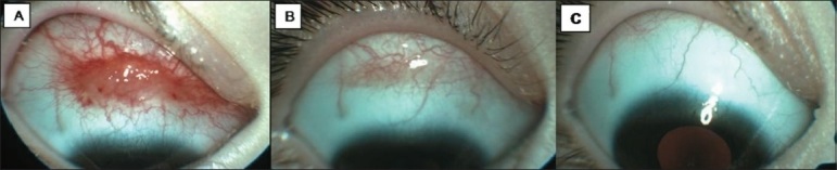
A 3-year-old girl presented with a hemorrhagic conjunctival lesion in the right eye. The medical history revealed premature cessation of breast feeding, intolerance to the ingestion of baby foods, anorexia, and abdominal distention. Prior to her referral, endoscopic small intestinal biopsy had been carried out under general anesthesia with a possible diagnosis of Celiac Disease (CD). Her parents did not want their child to undergo general anesthesia for the second time for the excisional biopsy. We decided to follow the patient until all systemic investigations were concluded. In evaluation, the case was diagnosed with CD and the conjunctival tumor showed complete regression during gluten-free dietary treatment. The clinical fleshy appearance of the lesion with spider-like vascular extensions and subconjunctival hemorrhagic spots, possible association with an acquired immune system dysfunction due to CD, and spontaneous regression by a gluten-free diet led us to make a presumed diagnosis of conjunctival Kaposi sarcoma.
Celiac disease (CD) is an autoimmune gluten-induced enteropathy that typically causes intestinal malabsorption syndrome in childhood. The clinical course may be complicated by extra-intestinal immunological disorders such as dermatitis herpetiformis, cutaneous vasculitis, polyarteritis, glomerulonephritis, chronic liver disease, polymyositis, and myocarditis.[1] Although rare, ocular involvement has been reported as well.[2–4] These include immune-complex mediated uveitis,[2] ocular myopathy,[3] and Vogt-Koyanagi-Harada disease.[4] We report herein a unique case with a very unusual conjunctival tumor that showed complete regression during dietary treatment of CD.
Case Report
A 3-year-old girl presented with a complaint of hemorrhagic tear episodes of 3 months duration in the right eye. Her parents stated that the reddish conjunctival lesion was detected elsewhere one month ago and was unresponsive to topical steroids, and hence, excisional biopsy was suggested. The patient was referred to our clinic to get a second opinion.
Her past medical history revealed premature cessation of breast feeding, intolerance to the ingestion of baby foods, anorexia, and abdominal distention since 2 months. Her weight and height percentiles were subnormal compared to her age group. From 26 months of age, she had recurrent serous otitis media treated with systemic antibiotics. However, the primary etiology could not be determined by her pediatrician in the first 3 years of life.
Our initial visit showed that the visual acuities were 20/20 in both eyes. Slit-lamp examination of the right eye revealed a reddish, elevated, and highly vascular spider-like lesion on the superior bulbar conjunctiva, measuring 12×4×2 mm [Fig. 1A]. Our presumed diagnosis was conjunctival Kaposi sarcoma (KS). Prior to her referral, endoscopic small intestinal biopsy had been carried out under general anesthesia with a possible diagnosis of CD in another hospital. Therefore, her parents did not want their child to undergo general anesthesia for the second time for the excisional biopsy. We decided to follow the patient without any intervention until all systemic investigations were concluded.